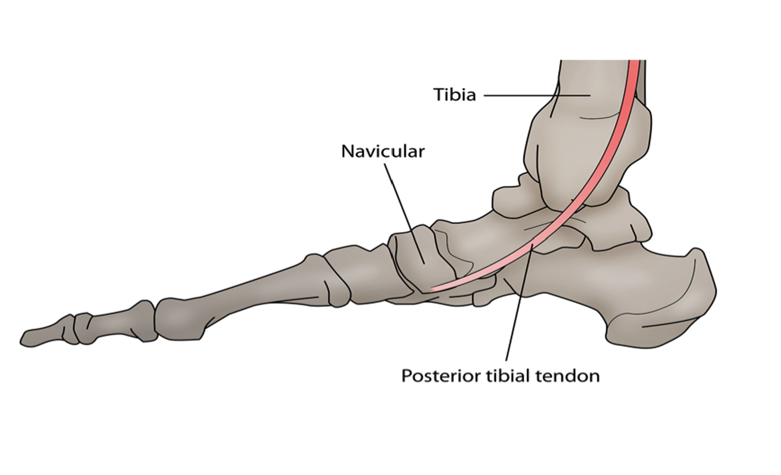
The posterior tibial tendon, located in the back of the leg beneath the calf muscle, plays a crucial role in foot function and structure.
Posterior Tibial Tendon Dysfunction (PTTD) is a condition where the tendon becomes degenerated, thickened, and structurally compromised, leading to muscle weakness and progressive flattening of the arch.
PTTD can be diagnosed through a medical history and a full biomechanical assessment, and imaging techniques such as X-ray and ultrasound.
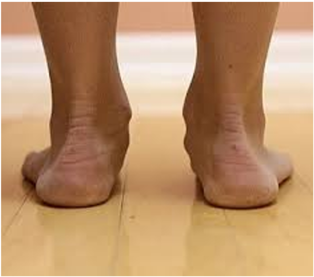
Flat feet or pes planus, which cause excessive pronation, are the most common cause of PTTD.
Treatment options include orthotics, exercises, footwear, strapping and supportive braces, and K-Laser therapy.
Learn about the tibialis posterior muscle and what it does.
The tibialis posterior is a muscle originating from the back of the leg found beneath the calf muscle.
The muscle is small in size and is positioned alongside other deep muscles of the leg such as the extensor digitorum longus and extensor hallucis longus muscles.
Despite the seemingly small muscle belly of the tibialis posterior muscle, it has one of the most important roles in foot function and structure. Tibialis posterior assists in plantarflexion of the foot (allowing you to stand on your toes). Functioning as the primary supinator of the foot, it’s job is to hold the arch up and stop the foot from overpronating or rolling in too much during walking and running.
When this muscle is injured or not working as well as it should be, this can be frustrating.
What is the difference between tibialis posterior tendonitis and posterior tibial tendon dysfunction?
Tibialis posterior tendonitis is inflammation of the tendon, this will often be the result of an injury tendon or an increased workload the tendon is placed under. The workload the tendon is put under can be increased by the structure of the foot and walking pattern. Tibialis posterior tendonitis can lead to tendon dysfunction and is the next more serious progression of the condition that happens if the tendonitis is not sufficiently treated in the early stages of the disease.
PTTD occurs when the tendon progresses from the inflammatory stage to becoming degenerated, thickened and the actual structure of the tendon has become compromised. At this stage, the tendon no longer functions as it once did, resulting in muscle weakness and a progressive flattening of the arch.
How do I know if I have PTTD, what are the symptoms?
Some of the symptoms you may experience if you have PTTD may include:
If we are concerned that you may have PTTD, we will put you through a comprehensive medical assessment to arrive at a diagnosis. This will include:
Taking a thorough medical history:
We will work with you to get a clear picture of your precise medical history. There are often clues in your past that help us work out what is happening in the present. Information such as previous foot or lower limb injuries, the types of activities you take part in and pain you may have experienced could all provide helpful information.
A full Biomechanical assessment:
This will include a static (standing) assessment of your feet and ankle looking at the posture and position of your feet.
We will also perform a number of clinical tests specific to this PTTD that establish muscle strength, joint range of motion and the alignment of the joint axis within your feet. The joint axis has a large impact on how your feet function when walking and running.
We also perform a dynamic assessment where we have you walk and or run on a treadmill and capture this on video so we can determine the best treatment approach for you.
Imaging
We mainly use x-ray and ultrasound imaging to assist us in accurately diagnosing PTTD. These images are very important in helping us determine the severity of the condition and whether other structures are involved or damaged.
What causes posterior tibial tendon dysfunction?
The most common cause of PTTD is flat feet or feet that collapse and pronate heavily when walking. These types of feet are known medically as pes planus. Tibialis Posterior is the main supinator of the foot (supination is the opposite movement to pronation) as such in a pes planus foot, where pronation is excessive, tibialis posterior is forced to work harder to supinate the foot. This extra workload can result in fatigue and damage to the tendon and in more extreme cases result in fraying and even partial or full thickness tearing of the tendon.
Repeated loading through walking and running without support from the right footwear and insoles can be the catalyst for the arch collapse we often see with this condition. When this condition really deteriorates you can experience additional damage to the ankle and midfoot with damage to the arch ligament (spring ligament) and chronic arthritis forming in around the midfoot as well.
How is PTTD treated?
Treatment available for tibialis posterior dysfunction are.
Orthotics
Orthotics can help to correct the position of your foot and improve your arch by providing support and guidance for your feet. If you’re in the early stages of this kind of injury/damage, the use of orthotics may allow for you to avoid bigger problems in the future. We offer custom orthotics here at Hurst Podiatry.
Exercises
Programs designed to help create mobility and strength can help overcome some of the weaknesses and deficiencies that lead to PTTD in the first place. We can help build such a program through you through our digital physitrack program.
Footwear
The right shoes can make all the difference when it comes to the health of your feet, lower limbs and body overall. Shoes that have little to no support end up resulting in you putting more pressure on the tibialis posterior tendon. The right shoes can prevent this from occurring and a whole range of other conditions.
Strapping and Supportive braces
There are a number of taping and strapping techniques we can employ that help support the plantar fascia and reduce stress on the area to reduce symptoms.
K-Laser therapy
K laser therapy involves using photobiomodulation via UV frequencies on chromophores(specfic body cells) to create positive healing changes like stimulating blood flow, increasing collagen production, stimulating and boost cell production. There are only a handful of K lasers available in the state and we are lucky enough to have one at our clinics. You can learn more about K laser here.
Shockwave therapy
Shockwave therapy involves using a therapy unit on the site of the injury to stimulate new healing and blood flow. You can learn more about its benefits here.
Surgery
If the tibialis posterior tendon is severely damaged, such as having been torn or the bones of the area have been damaged, surgery could be the best solution. Your podiatrist will work with you to ensure you understand your options before any type of surgery is recommended.
How long will it take to get better?
This will be determined mainly by the severity of the injury with more chronic cases taking longer to go away than more minor cases. Most cases of PTTP will be solved with conservative treatment within 4-10 weeks though with a strong management plan. If it is taking much longer than this, we often consider a surgical option.
Visit our podiatry clinic in Melbourne
If you’re looking for a podiatrist to check out your heel spur or any other foot condition, we’re here to help you. We’re in Melbourne’s Eastern suburbs, with clinics located in Croydon, Healesville, Kilsyth and Mooroolbark. Our highly experienced team is here to help you take care of your feet today.
I was surfing through the internet for a while just to get in-depth information about the topic you wrote about and it really helped me to know the useful information. Keep sharing such blogs further as well. And kindly let me know how can I subscribe to the Newsletter. Thanks.
Hi Tim! we’re thrilled this has been helpful. We don’t currently have a newsletter in place but do have plans of adding one to our offerings shortly. We do post blogs here very regularly though so keep your eyes peeled! Thanks again